Fetoscopic Surgery
"It sounded like we were looking at brain damage, feeding tubes, a breathing tube, a wheelchair, just a bad quality of life [from an ultrasound diagnosis at 13 weeks gestation]."
"It's not done by any means, but I definitely feel it's [fetoscopic surgery] the right thing for us. I can't imagine going on further in the pregnancy not knowing every day what damage is being done and if he's getting worse."
"It's such a relief to move forward."
Mother, Lexi Royer, 28, Texas Children's Hospital, Houston
| Part of the spinal cord and spinal nerves, usually encased in a sac, protrude through an opening in the back and is exposed to the amniotic fluid. The brainstem (hindbrain) descends, or herniates, into the spinal canal in the neck and blocks the flow of cerebrospinal fluid. This can cause a damaging buildup of fluid in the brain called hydrocephalus |
Although fetal surgery in the repair of spina bifida has been performed since the 1990s, an entirely new technique has been pioneered, an experimental technique that has some experts in the field doubtful whether it is completely safe. At three to four weeks of pregnancy tissue forming the spinal column in a developing fetus is meant to fold into a tube; with the onset of spina bifida the tube fails to close, as it should. Why this occurs is not fully understood, but a vitamin B folic acid deficiency is known to be involved.
Mrs. Royer and her husband Joshuwa who live in San Diego, were informed that their developing child had a defect, a severe one where the brain stem was pulled into the spinal column, the result of spina bifida. At 24 weeks their fetus weighed under one kilo and surgery was being undertaken for his severe form of spina bifida. The Royers knew that this condition meant that children are unable to walk and fluid builds up in the brain, along with other complications.
Pediatric neurosurgeon Dr. William Whitehead and the obstetrician/gynecologist-in-chief at Texas Children's Hospital prepared to operate. And what unfolded was the experimental technique where the surgeons made a wide incision in the mother's lower abdomen and then with great care, lifted her uterus, still internally attached, free of her abdomen. Two tiny slits were made in the uterus, one of which was to insert a "fetoscope" (telescopic camera, light, and grasping tool). The second slit was meant for the use of other, miniaturized surgical instruments.
In 2011 a study was published concluding that prenatal surgery was a superior technique with a more promising outlook in comparison to operating after birth. The need for a shunt was halved in those children who underwent surgery before birth, and the number of those who were enabled to walk independently rose to 40 percent from the 20 percent success seen in post-birth surgery. "The percent who benefit, I wish it was higher" said Dr. Whitehead.
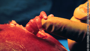 |
| During surgery on a 22-week-old fetus at Vanderbilt, Dr. Joseph Bruner demonstrates size by comparing the fetus' hand to his index fingertip. |
In standard prenatal surgery for spina bifida, the woman's abdomen is cut open and then the uterus to enable the fetus to be operated on. In the experimental approach Dr. Belfort opened Mrs. Royer's lower abdomen. The surgeons operating on the fetus through the small slit in the uterus raised out of and above the abdomen, pulled skin and membranes to cover the naked spinal cord, then sewed them, sealing out amniotic fluid, in a three-hour procedure.
This was not the first time their experimental technique had been used, bur the 28th time. The technique is not universally accepted, with some doctors warning that carbon dioxide pumped into the uterus to expand it and enable the use of the tiny surgical instruments in the delicate operation, might have a harmful effect on the fetus. Dr. Belfort rejects that concern, observing that there has been no evidence of harm done, though the passage of time will be more explicit.
Labels: Bioscience, Research, Science, Spina Bifida, Surgery
posted by Pieface @ 6:13 PM
![]()
![]()

0 Comments:
Post a Comment
<< Home