Bioprinting Brain Tumours
"I would put 1,000 cells each on two petri dishes and treat one with a chemotherapeutic agent, The next day, or three days later, I expect to see the treated cells to be reduced to 10% of the original cells, while the control will continue to multiply every day."“"It’s outrageous! And it means that something is wrong. I started to wonder. I got to the point of thinking that maybe we were working with the wrong cancer model.""We found that this protein is responsible for a failure in the microglia, causing them to support rather than attack the deadly cancer cells, helping the cancer spread, However, we identified the protein in tumors removed during surgery, but not in glioblastoma cells grown on 2D plastic petri dishes in our lab.""Each model is printed in a bioreactor we have designed in the lab using a hydrogel sampled and reproduced from the extracellular matrix taken from the patient, thereby simulating the tissue itself.""The physical and mechanical properties of the brain are different from those of other organs, like the skin, breast or bone. Breast tissue consists mostly of fat; bone tissue is mostly calcium. Each tissue has its own properties, which affect the behavior of cancer cells and how they respond to medications.""First, we tested a substance that inhibited the protein we had recently discovered, P-Selectin, in glioblastoma cell cultures grown on 2D petri dishes, and found no difference in cell division and migration between the treated cells and the control cells which received no treatment. In contrast, in both animal models and in the 3D-bioprinted models, which overexpress the protein, we were able to delay the growth and invasion of glioblastoma by blocking the P-Selectin protein.""We had a lot of difficulties and challenges on the way.""If we take a sample from a patient’s tissue, together with its extracellular matrix, we can 3D bioprint from this sample 100 tiny tumors and test many different drugs in various combinations to discover the optimal treatment for this specific tumor, Alternatively, we can test numerous compounds on a 3D-bioprinted tumor and decide which is most promising for further development and investment as a potential drug.""But perhaps the most exciting aspect is finding novel druggable target proteins and genes in cancer cells – a very difficult task when the tumor is inside the brain of a human patient or model animal.”"We have about two weeks [to] test all the different therapies that we would like to evaluate [on] that specific tumour, and get back with an answer -- which treatment is predicted to be the best fit."Professor Ronit Satchi-Fainaro, Tel Aviv University, Israel
 and pericytes (cyan). The blood vessels are surrounded with a brain-mimicking tissue composed of gliblastoma cells (blue) and the brain microenvironment cells (green).") |
Microscopic image of the 3D-bioprinted
glioblastoma model. The bioprinted blood vessels are covered with
endothelial cells (red) and pericytes (cyan). The blood vessels are
surrounded with a brain-mimicking tissue composed of gliblastoma cells
(blue) and the brain microenvironment cells (green). (photo credit: TEL AVIV UNIVERSITY)
|
"The more physiological mimicry you create, the better prediction you get in terms of how drug treatments will work on the actual tumour in the patient's body."Ofra Benny, Hebrew University of Jerusalem
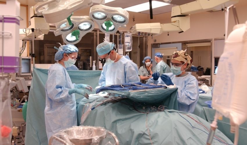
New research out of Israel, just recently published in the journal Science Advances, chronicled an advance in the treatment of brain cancer. Researchers made use of brain cancer patients' specific cells in a form of 3D printing material to produce a model of the cancer tumour which would enable them to test the efficacy of various possible treatments to assess them before making use of them within the patient's body.
A portion of the tumour would be extracted from a patient's brain who suffered from glioblastoma -- an aggressive cancer whose outcome is never too assured given its poor prognosis -- to make use of the extracted portion in printing a model matching MRI scans, explained Professor Satchi-Fainaro, leading the research team at Tel Aviv University.
Bioprinting: has been used in previous research simulating cancer environments, but according to the Tel Aviv University researchers' claim, they represent the first to print "viable" tumours. Should the printed tumour shrink, or be seen to lower metabolic activity against control groups, the treatment used is considered promising.
In planning for surgery 3D printed tumour models have been printed. Recent innovations, however have sought to focus on bioprinting with the use of live cells as a bio-guide to building up layers. According to Ofra Benny of the Hebrew University of Jerusalem, who herself leads research that is similar, the use of a patient's own cells to develop 3D tumour models could be "a game-changer" in the understanding and accuracy and success in treating these most difficult of tumours.
: Eilam Yeini, Prof. Satchi-Fainaro and Lena Neufeld. (credit: TEL AVIV UNIVERSITY)") |
| (Left to right): Eilam Yeini, Prof. Satchi-Fainaro and Lena Neufeld. (credit: TEL AVIV UNIVERSITY) |
Labels: Bioprinting, Brain Tumours, Research, Treatment
posted by Pieface @ 8:01 PM
![]()
![]()

0 Comments:
Post a Comment
<< Home