Ruminations

Blog dedicated primarily to randomly selected news items; comments reflecting personal perceptions

Wednesday, December 15, 2021

The Link Between Asthma and Brain Tumours

"Of course, we're not going to start inducing asthma in anyone; asthma can be a lethal disease. But what if we could trick the T cells into thinking they're asthma T cells when they enter the brain, so they no longer support brain tumour formation and growth?"
"These findings open the door to new kinds of therapies targeting T cells and their interactions with cells in the brain."
"The most exciting part of this is that it shows that there is a normal communication between T cells in the body and the cells in the brain that support optic pathway glioma formation and growth."
"The next step for us is to see whether this is also true for other kinds of brain tumours. We're also investigating the role of eczema and early-childhood infections, because they both involve T cells."
David Gutmann, researcher, expert on neurofibromatosis, professor of genetics, of neurosurgery and paediatrics, Washington University School of Medicine, St.Louis 

Woman with asthma using inhaler
(bobex73 - stock.adobe.com)
 
Insatiable curiosity teamed with the methodology and collaboration between inquisitive scientific minds often represent the emerging blueprint for discovery of new potential therapies in solving human ills that often threaten lives. Researchers at Washington University School of Medicine in St.Louis discovered that when mice develop asthma, their T cells activate in such a manner that induces inflammation in the lungs, yet prevents growth of tumours in the brain.

In essence, the research team may just have happened upon a new method of designing a response to brain tumors after discovering just why it is that the peculiar and unproven previously-known connection between asthma sufferers being less likely than others to go on to develop this insidious form of cancer. Published in the journal Nature Communications, the study explored the relationship, mysterious as it is, between inflammation diseases and brain tumours.

These two conditions so very different from one another, long puzzled the medical community in a possible connection, despite which new research has led to the discovery that T cells in the body may hold the answer to a question that has long mystified scientists. When mice develop asthma, their T cells activate to induce inflammation in the lungs, an effect that also has the effect of preventing growth of tumours in the brain. 

It was first postulated 15 years ago that inflammatory diseases have a relation to reduced risk of brain tumours, based largely on epidemiologic observations. No obvious link presented, however, linking the two conditions, leaving many researchers in doubt that an association actually was present. Dr. Gutmann, senior scientist in the study, noticed an inverse relationship between the two conditions some five years earlier through study of neurofibromatosis; a set of complex genetic disorders leading to the growth of tumours on nerves in the brain and throughout the body.

Uncertain how to link to a connection, more recent research taking place in his laboratory identified the key role that immune cells play in formation of tumours on the optic pathways of children with type 1 NF (neurofibromatosis). A collaboration resulted with Jit Chatterjee, post-doctoral researcher who was in the process of diagnosing the underlying mechanism at work. Dr.Chatterjee exposed mice that were engineered to carry a mutation in the NF1 genes to result in tumours on their optic nerves.

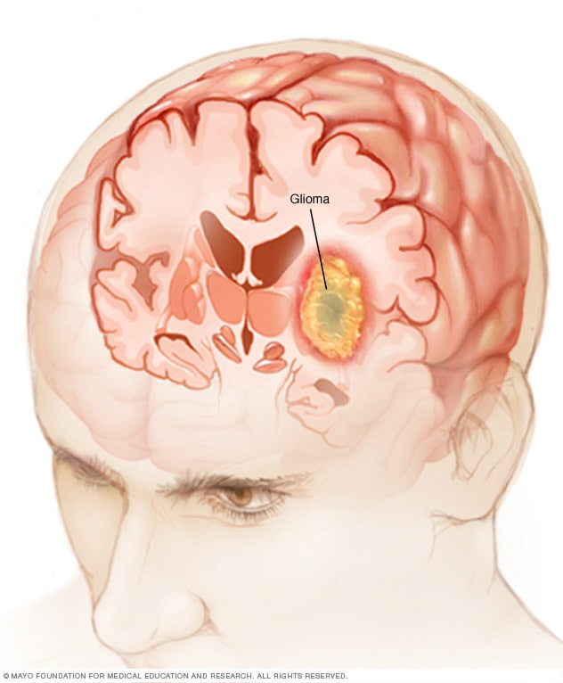
Glioma
These young mice in their formative period of growth were exposed to irritants known to be the cause of asthma, administering a salt-water solution to a control group of mice. The mice that developed asthma turned out not to experience tumour growth on their optic nerves. Then it was revealed through further study that the T cells of these mice were behaving differently after developing asthma, secreting a protein named decorin, known to act on and irritate the lining of the brain airways.

The protein blocked the activation of immune cells called microglia, that promote tumour growth. "As we understand this communication between T cells and the cells that promote brain tumors better, we’ll start finding more opportunities to develop clever therapeutics to intervene in the process", explained Dr. Gutmann.


Labels: , ,

Tuesday, August 24, 2021

Bioprinting Brain Tumours


"I would put 1,000 cells each on two petri dishes and treat one with a chemotherapeutic agent, The next day, or three days later, I expect to see the treated cells to be reduced to 10% of the original cells, while the control will continue to multiply every day."
“"It’s outrageous! And it means that something is wrong. I started to wonder. I got to the point of thinking that maybe we were working with the wrong cancer model."
"We found that this protein is responsible for a failure in the microglia, causing them to support rather than attack the deadly cancer cells, helping the cancer spread, However, we identified the protein in tumors removed during surgery, but not in glioblastoma cells grown on 2D plastic petri dishes in our lab."
"Each model is printed in a bioreactor we have designed in the lab using a hydrogel sampled and reproduced from the extracellular matrix taken from the patient, thereby simulating the tissue itself."
"The physical and mechanical properties of the brain are different from those of other organs, like the skin, breast or bone. Breast tissue consists mostly of fat; bone tissue is mostly calcium. Each tissue has its own properties, which affect the behavior of cancer cells and how they respond to medications."
"First, we tested a substance that inhibited the protein we had recently discovered, P-Selectin, in glioblastoma cell cultures grown on 2D petri dishes, and found no difference in cell division and migration between the treated cells and the control cells which received no treatment. In contrast, in both animal models and in the 3D-bioprinted models, which overexpress the protein, we were able to delay the growth and invasion of glioblastoma by blocking the P-Selectin protein."
"We had a lot of difficulties and challenges on the way."
"If we take a sample from a patient’s tissue, together with its extracellular matrix, we can 3D bioprint from this sample 100 tiny tumors and test many different drugs in various combinations to discover the optimal treatment for this specific tumor, Alternatively, we can test numerous compounds on a 3D-bioprinted tumor and decide which is most promising for further development and investment as a potential drug."
"But perhaps the most exciting aspect is finding novel druggable target proteins and genes in cancer cells – a very difficult task when the tumor is inside the brain of a human patient or model animal.”
"We have about two weeks [to] test all the different therapies that we would like to evaluate [on] that specific tumour, and get back with an answer -- which treatment is predicted to be the best fit."
Professor Ronit Satchi-Fainaro, Tel Aviv University, Israel
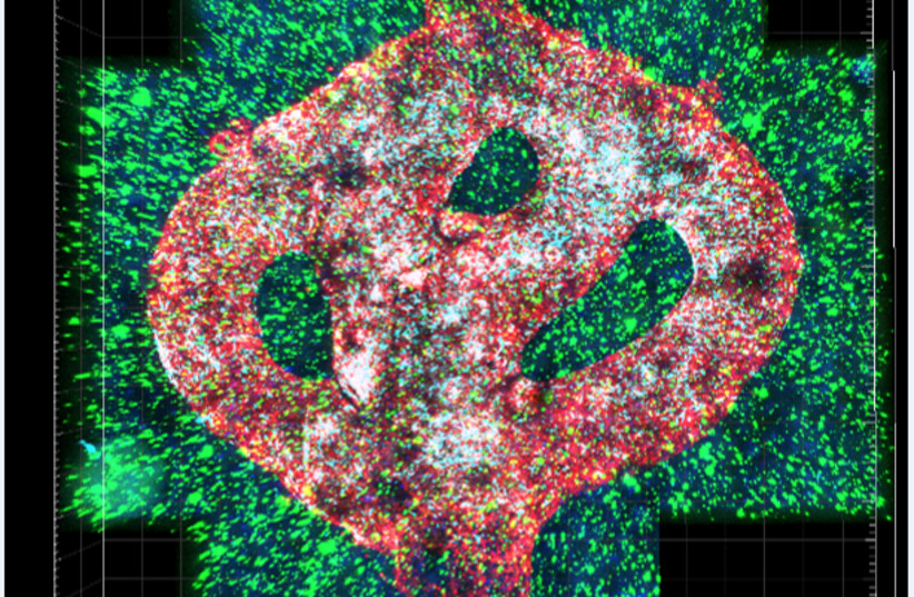
 Microscopic image of the 3D-bioprinted glioblastoma model. The bioprinted blood vessels are covered with endothelial cells (red) and pericytes (cyan). The blood vessels are surrounded with a brain-mimicking tissue composed of gliblastoma cells (blue) and the brain microenvironment cells (green).  (photo credit: TEL AVIV UNIVERSITY)
Microscopic image of the 3D-bioprinted glioblastoma model. The bioprinted blood vessels are covered with endothelial cells (red) and pericytes (cyan). The blood vessels are surrounded with a brain-mimicking tissue composed of gliblastoma cells (blue) and the brain microenvironment cells (green).    (photo credit: TEL AVIV UNIVERSITY)

"The more physiological mimicry you create, the better prediction you get in terms of how drug treatments will work on the actual tumour in the patient's body."
Ofra Benny, Hebrew University of Jerusalem
New research out of Israel, just recently published in the journal Science Advances, chronicled an advance in the treatment of brain cancer. Researchers made use of brain cancer patients' specific cells in a form of 3D printing material to produce a model of the cancer tumour which would enable them to test the efficacy of various possible treatments to assess them before making use of them within the patient's body.

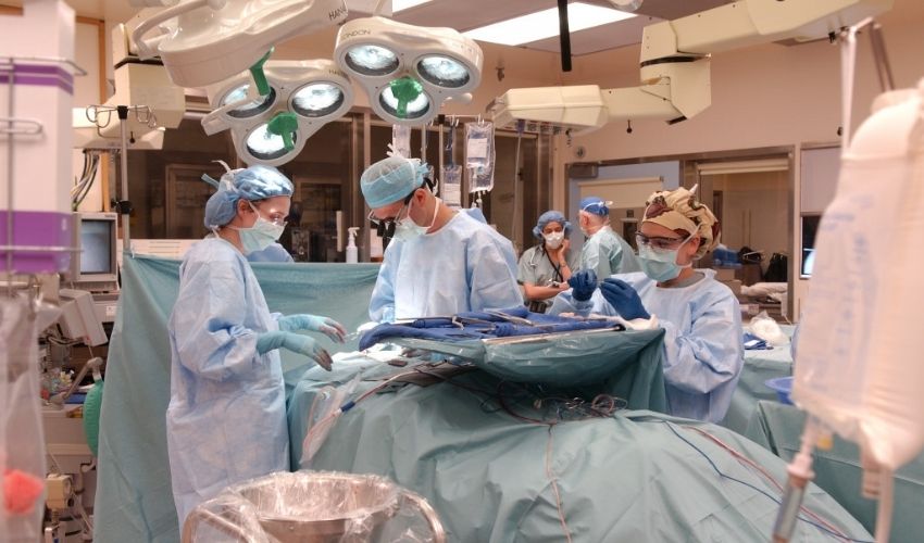
A portion of the tumour would be extracted from a patient's brain who suffered from glioblastoma -- an aggressive cancer whose outcome is never too assured given its poor prognosis -- to make use of the extracted portion in printing a model matching MRI scans, explained Professor Satchi-Fainaro, leading the research team at Tel Aviv University.

Bioprinting: has been used in previous research simulating cancer environments, but according to the Tel Aviv University researchers' claim, they represent the first to print "viable" tumours. Should the printed tumour shrink, or be seen to lower metabolic activity against control groups, the treatment used is considered promising. 
 
In planning for surgery 3D printed tumour models have been printed. Recent innovations,  however have sought to focus on bioprinting with the use of live cells as a bio-guide to building up layers. According to Ofra Benny of the Hebrew University of Jerusalem, who herself leads research that is similar, the use of a patient's own cells to develop 3D tumour models could be "a game-changer" in the understanding and accuracy and success in treating these most difficult of tumours.

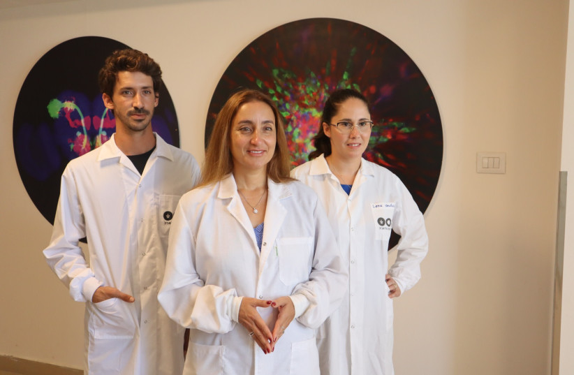
 (Left to right): Eilam Yeini, Prof. Satchi-Fainaro and Lena Neufeld. (credit: TEL AVIV UNIVERSITY)
(Left to right): Eilam Yeini, Prof. Satchi-Fainaro and Lena Neufeld. (credit: TEL AVIV UNIVERSITY)


Labels: , , ,

 
()() Follow @rheytah Tweet