Ruminations

Blog dedicated primarily to randomly selected news items; comments reflecting personal perceptions

Monday, December 27, 2021

In For The Long Haul With COVID

"This is remarkably important work. For a long time now, we have been scratching our heads and asking why long COVID seems to affect so many organ systems."
"This paper sheds some light, and may help explain why long COVID can occur even in people who had mild or asymptomatic acute disease."
Zyad Al-Aly, director, clinical epidemiology centre, Veterans Affairs St.Louis Health Care System, Missouri
 
"We don't fully understand long COVID, but these changes could explain ongoing symptoms."
"[The research] provides a warning about being blase about mass infection in children and adults."
"We don't yet know what burden of chronic illness will remain in years to come. Will we see young-onset cardiac failure in survivors, or early onset dementia?"
"These are unanswered questions which call for a precautionary public health approach to mitigation of the spread of the virus."
Raina MacIntyre, professor of global biosecurity, University of New South Wales, Sydney

"Our results collectively show that while the highest burden of SARS-CoV-2 is in the airways and lung, the virus can disseminate early during infection and infect cells throughout the entire body, including widely throughout the brain."
NIH Study authors
Coronavirus Can Persist For Months In Heart, Brain: Study
 
A new study concludes that the coronavirus causing COVID-19 has the ability to spread within days from the airways to the heart, brain and just about every bodily organ system, in addition to which it may persist for months. The study authors describe their work as the most comprehensive analysis to the present of the SARS-CoV-2 virus's widespread distribution and duration in both body and brain. 
 
Scientists at the U.S. National Institutes of Health discovered that the pathogen has the capacity to replicate in human cells beyond the respiratory tract.

The manuscript, pre-released on line, is currently under review preparatory to publication in the journal Nature; its results spell out delayed viral clearance as a possible contributor to the pernicious symptoms stubbornly wracking the minds and bodies of long-COVID sufferers. Knowing how the virus is able to persist, as well as the body's response to a viral reservoir has the potential to aid in improving care for those sufferers.

The investigative discoveries and the techniques used are yet to be reviewed by independent scientists, and relate to data gathered from fatal COVID cases, and not from people suffering with long COVID or "post-acute sequelae of SARS-CoV-2", the authors point out. Numerous earlier studies provide evidence both for and against the possibility that the coronavirus has the propensity to infect cells outside the airways and lungs.
 
Undertaken at the National Institutes of Health in Bethesda, Maryland, the research is based on sampling extensively, and analysis of tissues reserved during autopsies on 44 American patients who failed to survive their encounter with the coronavirus. Daniel Chertow, who heads the NIH emerging pathogen section, wrote that the burden of infection outside the respiratory tract and the time it takes to clear the virus from infected tissues, particularly in the brain, fail to be characterized.

Persistent SARS-CoV-2 RNA was detected by the study group in multiple parts of the body, as well as throughout the brain, for up to 230 days after symptom onset, which may represent infection with defective virus particles, seen as well in persistent infection with the measles virus. The NIH team's post-mortem tissue collection was comprehensive, occurring within about a day of the patients' demise.

A variety of tissue preservation techniques to detect and quantify viral levels was also used, along with growing the virus collected from multiple tissues, including lung, heart, small intestine and adrenal gland. Pathological data can be drawn from the study supporting findings of previous research indicating, as an example, that SARS-CoV-2 directly destroys heart muscle cells, and those who survive an infection suffer cognitive deficits.

Infection of the pulmonary system may result in an early "viremic" phase where the virus is seeded throughout the body, including in patients experiencing mild or no symptoms, posit the NIH researchers. An example given was that of a juvenile included in the autopsy study who was presumed to have died from unrelated seizure complications, suggesting infected children with mild COVID-19 onset are also able to experience systemic infection.
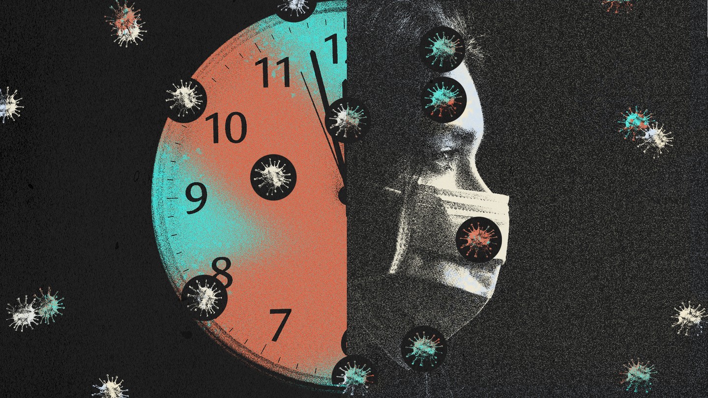
A masked face next to a clock, surrounded by coronaviruses
Getty -- The Atlantic

Labels: , , , , , ,

Monday, January 06, 2020

Hope For Alzheimer's


"Photobiomodulation introduces the therapeutic effect of light into our brain. It triggers the body to restore its natural balance or homeostasis. When we do that, we call upon the body's innate ability to heal."
"We have a much bigger ambition than the drug trials. Drug developers are mainly either seeking to slow the mental decline in diagnosed cases, or to prevent the onset of Alzheimer's disease by intervening at the pre-symptomatic stage."
"Based on early data, we are confident of seeing some measure of recovery in the symptoms, not just a slowdown in the rate of decline, even in moderate to severe cases."
Dr.Lew Lim, CEO, Vielight Inc.
The Vielight Neuro RX Gamma device in use.


In Canada, an estimated half-million Canadians are living currently with Alzheimer's disease. In the United States that number is around 5.8 million. New cases diagnosed every year number around 25,000 in Canada, a total number expected to rise in Canada by 66 percent for a total of 937,000 people by the year 2031. Since the U.S. has about ten times the population of Canada, the diagnosed incidence of Alzheimer's onset is quite similar in each population base. Worldwide the number of those living with Alzheimer's is around 50 million, although it would appear that only one in four sufferers has been diagnosed, according to Alzheimer's Disease International.
"In spite of substantial efforts to establish effective treatments for Alzheimer’s disease, progress has been, unfortunately, limited and a cure does not yet exist."
"Although not conventional as an Alzheimer’s disease solution, we look forward to investigating the potential of Vielight’s technology in the treatment of this difficult disease based on promising early evidence."
Dr. Corinne Fischer, principal investigator, St.Michael's Hospital, Toronto
The light is directed at specific areas of the brain known to be damaged in Alzherimer's
The light is directed at specific areas of the brain known to be damaged in Alzherimer's Credit: Vielight
Currently, there is no treatment for Alzheimer's to cure the condition or to prevent its onset, although drug treatment has succeeded in slowing its progress in a minor way. The drug that appeared to give the most hope for the future, Biogen's aducanumah has been seen to slow dementia onset, but that appears the extent of its efficacy. No new drugs have been developed in almost two decades. Scientists have by and large surrendered their hope of reversing brain damage, once it has occurred.

On the near horizon, however, another treatment has recently emerged and has undergone initial clinical trials.

Resulting in the first major trial to determine whether light therapy might turn out to be beneficial for dementia. Early findings have astounded the scientific world, when people have been seen to regain memory, their reading and writing capability and have realized restored orientation, all negatively impacted and increasingly diminished by Alzheimer's. Should the trial ultimately be deemed successful it would represent the first treatment enabling the reversal of the disease's steady brain and body decline.
Multiple mechanisms for PBM in brain

The focus of the trial is a device named the Neuro RX Gamma headset, developed by Vielight, a biotech company based in Toronto. A process called "photobiomodulation" has been devised where parts of the brain known to be damaged in dementia are exposed to pulses of near-infrared light. Using LEDs on a headset, the device fires 4 Hz gamma waves directly into the skull. The hippocampus, part of the brain connected to memory, sees additional light channeled up the patient's nose by use of a separate nasal clip.
dementia symptoms

A pilot trial to test the effects of home photobiomodulation (PBM) on cognitive and behavioral function, cerebral perfusion and functional connectivity in eight patients with dementia was administered in a home setting three times weekly with the use of the Vielight Neuro Gamma device pulsing at 40 Hz. Participants were assessed at 6 and 12 weeks. Following 12 weeks of use improvements were seen in all indices. Should the trial prove to be the success that early results appear to indicate, people suffering with Alzheimer's would be able to self-administer their treatments at home with use of the light-emitting device.
healthy brain versus alzheimers brain

Alzheimer’s is the most common cause of dementia among older adults. Dementia is the loss of cognitive functioning—thinking, remembering, and reasoning—and behavioral abilities to such an extent that it interferes with a person’s daily life and activities. Dementia ranges in severity from the mildest stage, when it is just beginning to affect a person’s functioning, to the most severe stage, when the person must depend completely on others for basic activities of daily living.
The causes of dementia can vary, depending on the types of brain changes that may be taking place. Other dementias include Lewy body dementia, frontotemporal disorders, and vascular dementia. It is common for people to have mixed dementia—a combination of two or more types of dementia. For example, some people have both Alzheimer’s disease and vascular dementia.
National Institute on Aging, U.S.

Labels: , , , , , , ,

Saturday, November 30, 2019

Mind Your Brain

"So now we know that pleasure is also encoded by opioids."
"The next time you're hungry and you see the McDonald's arches, that's leading to dopamine release. So it's predicting you're going to get reinforced or rewarded. But dopamine can also predict negative things."
"So if we're eating broccoli it's 10 units of dopamine. But when we're smoking crack cocaine, it's like 10,000. That's why for the average human seeking pleasure how can broccoli, or even brownies, compete with crack cocaine?"
"When you're activating this reward circuit, you're kind of tipping the balance up, and your brain is going to try to bring it down. It engages these compensatory responses."
"What you're feeling the next morning is the exact opposite of how you felt when you were on the stimulus. This would happen with any one of us if we were recreationally using."
"When you're using, you're activating all the circuits. And the next day, or the next week if you have a binge weekend, the circuits are kind of resetting the balance."
Dr. Kim Hellemans, chair, Department of Neuroscience, Carleton University
Mother talking to adolescent daughter.
MedicineNet.com

When humans come abreast of things required for survival, the brain releases a "reward circuit", as part of our evolutionary preparedness for the future of humankind. In primordial times when a successful hunt concluded that would ensure survival in the short term, brain circuits released a cascade of signals encoding pleasure, informing the memory to pay attention to where the successful event took place, so at a later time hunters could return with the expectation of another successful hunt.

Phenobarbital, Mebaral, Seconal, and Nembutal.

The hippocampus was engaged to inform the hunters where they were located, the nucleus accumbens lectured the mind how pleasurable the event had been with its expectations of a good tasting meal that would assure the dawning of another day, while the prefrontal cortex produced a mental note to return to the scene of success and pleasure. The brain is adept through long practise in its evolutionary trajectory in producing dopamine and releasing it to those brain areas alerting humans to survival techniques ensuring longevity.

Researchers have also discovered that the human brain released endorphins: opiates. The ingrained rewards system becomes engaged when we consume a substance such as cocaine, alcohol, nicotine, cannabis or opioids. That reward system becomes engaged in a superphysiological state, enhancing the experience of pleasure directly related to those substances. The complication is that the brain also is geared toward the maintenance of a steady balance (homeostatsis), and so engages in compensatory responses.
1130 opioids brain  Getty image brain scan Getty Images

Responses such as making it impossible for the dopamine to reach its reward potential by pulling the dopamine receptors from their location in the membrane even while  hormones that trigger stress are released, comprising the anti-reward circuits. Once the drug is metabolized and excreted, the high worn away, the compensatory responses are left; in the form of a 'hangover'. But in people gripped with a substance addiction where studies suggest early childhood trauma, the brain's circuits are rewired to blunt the reward baseline.

"That might have to do with depression or mental health, but they have this decreased reward baseline, and in some people, because of their history of trauma, they've got an overactive fear pathway." The stimuli that produce that good feeling in recreational users, makes traumatized people with addiction merely feel normal. Repeated use overrides the brain's stabilization efforts, setting a new, lower baseline to produce tolerance which people attempt to overwhelm through higher doses.

"Their baseline shifts, and people start feeling worse, even when they're not using. And they're trying to get to that level they did the first time they used, and they're never going to get there. This is 'chasing the dragon'."


"They'll say that they start using to feel good, and then they use just to feel normal, and then they're using to stay alive. Because what's happening is that when they're not using, the brain is saying 'Danger! Danger!". These fear pathways are skyrocketing, so you feel horrible and you feel scared. So when long-time users who are in withdrawal say they feel like they're dying, they actually feel like they're dying. Because all these fear pathways are going through the roof."

"All the decision-making in your prefrontal cortex is hijacked by addiction. When we're anxious, stressed, sad, a coping strategy can be alcohol, it can be opioids. And when you have an at-risk population -- people who may have experienced poverty, neglect, childhood trauma, systematic oppression due to the colour of their skin, their sexuality, their ethnic group -- this can certainly be the trigger that sets into motion a series of changes in the brain that may lead to addiction."

Labels: , , , , ,

Wednesday, June 20, 2018

Physical Exercise and the Aging Brain

"There is solid evidence to suggest that maintaining a regular exercise regimen can improve brain health."
"Each of these sessions [in an exercise program] gives you points that you rack up, and when we get to about 52 hours, then . . . we have this opportunity to reap the benefits of the exercise."
"You can think of it as exercise turning back the clock of age in your brain. We live in an era of instant gratification . . . but the encouraging thing about this is that it emphasizes the idea that you shouldn't be discouraged if you don't feel something in the first month. You just have to keep going."
"We are still learning about all the ways in which exercise changes our brain, and we are also all different, so identifying an ideal exercise dose remains a challenge. We have many more questions about exercise dose, and we will design further studies to follow up."
"We need to discuss this as a society because the global population is aging. By 2035, the scale is going to tip, and older adults [ages 65 and up] will be more plentiful than younger adults."
Joyce Gomes-Osman, post-doctoral research scholar, Beth Israel Deaconess Medical Center
"It's very encouraging that the evidence supports all sorts of different exercise interventions, not just aerobic, to improve thinking abilities. "
"The most stable improvements in thinking abilities were found in processing speed, both in healthy older adults and individuals with mild cognitive impairment." 
Alvaro Pascual-Leone, Chief of the Division of Cognitive Neurology, Director, Berenson-Allen Center for Noninvasive Brain Stimulation, Beth Israel Deaconess Medical Center
The researchers discovered that participants began experiencing cognitive improvements after working out for a total of 52 hours in approximately six months.
The researchers discovered that participants began experiencing cognitive improvements after working out for a total of 52 hours in approximately six months.  Mario Tama/Getty Images/File

In a joint study undertaken by researchers both at Harvard Medical School and Beth Israel it was found that any type of exercise, including walking, cycling, resistance training, yoga, weightlifting and so on is capable of having the astounding effect of improving cognitive and thinking skills in older people. Over 11,000 adults in their 70s were recruited to participate in the review, and its findings were published just recently in the journal Neurology Clinical Practise.

The researchers involved are convinced that even simply going out for a vigorous walk will help the elderly maintain their cognitive function. Lead author of the study, Joyce Gomes-Osman, has expressed her hope that the study findings will impress a significant number of people to encourage them that beginning an exercise plan and sticking to it, is not all that difficult, and the rewards make it well worth while for the effort.

After working out for a total of 52 hours within a six month period, study participants exhibited clear cognitive improvements, although researchers were quick to point out that noticeable benefits will vary from person to person, as well as the period of time it takes with a dedicated exercise program to begin experiencing those positively uplifting cognitive-improvement rewards. Though the benefits accruing from an exercise regimen accumulate over the long term, patience is required.

An attempt to accelerate the process would not necessarily turn out to be beneficial since it was determined that more intense exercise sessions of a shorter duration don't make the grade; in comparison to a steady exercise routine undertaken over a longer committal period. Aging while remaining mentally alert is a topic concerning greater numbers of people as peoples' longevity and expectations of remaining both physically and mentally alert consumes the public interest.

As we age, scientists have affirmed, our central nervous system slows down the production of new cells in our brain's hippocampus, affecting thinking and problem-solving. Still, according to researchers, regular commitment to endurance exercise is capable of stimulating the production of a protein called brain-derived neurotrophic factor, identified in a 2016 Harvard Medical School study.

That protein, over time "helps new neurons survive and thrive", noted Dr. Gomes-Osman. "Sometimes it's hard to translate these studies to people's lives, but I think we took a real important step in creating advice to translate results of evidence of exercise and brain health to a practical application that's applicable to everybody."

Labels: , , ,

Sunday, April 29, 2018

Preserving Brain Functionality

Abstract
Sedentary behavior associated with reduced medial temporal lobe thickness in middle-aged and older adults
Atrophy of the medial temporal lobe (MTL) occurs with aging, resulting in impaired episodic memory. Aerobic fitness is positively correlated with total hippocampal volume, a heavily studied memory-critical region within the MTL. However, research on associations between sedentary behavior and MTL subregion integrity is limited. Here we explore associations between thickness of the MTL and its subregions (namely CA1, CA23DG, fusiform gyrus, subiculum, parahippocampal, perirhinal and entorhinal cortex,), physical activity, and sedentary behavior. We assessed 35 non-demented middle-aged and older adults (25 women, 10 men; 45–75 years) using the International Physical Activity Questionnaire for older adults, which quantifies physical activity levels in MET-equivalent units and asks about the average number of hours spent sitting per day. All participants had high resolution MRI scans performed on a Siemens Allegra 3T MRI scanner, which allows for detailed investigation of the MTL. Controlling for age, total MTL thickness correlated inversely with hours of sitting/day (r = -0.37, p = 0.03). In MTL subregion analysis, parahippocampal (r = -0.45, p = 0.007), entorhinal (r = -0.33, p = 0.05) cortical and subiculum (r = -0.36, p = .04) thicknesses correlated inversely with hours of sitting/day. No significant correlations were observed between physical activity levels and MTL thickness. Though preliminary, our results suggest that more sedentary non-demented individuals have less MTL thickness. Future studies should include longitudinal analyses and explore mechanisms, as well as the efficacy of decreasing sedentary behaviors to reverse this association.
Neuroscience News logo


"Of course, we need larger samples and better ways to measure patterns of sedentary behavior. But if you're sitting for long periods of time, it seems that that factor — not physical activity — becomes the more harmful or more significant measure of your fitness. Even for people who are physically active, sitting a lot seems to be bad for your brain."
"[That's consistent with studies of sitting's effects on such health measures as heart disease, diabetes and mortality.] Now we're finding it in brain atrophy."
Prabha Siddarth, biostatistician, quantum chemist, UCLA


Too much sitting may thin the part of your brain that's important for memory, study suggests
Too much sitting, or other sedentary behavior, is associated with a thinning of a part of the brain important for memory, new research suggests. (Getty Images)
Those of the public interested in health studies will be aware that previous research has shown that sitting around for prolonged periods has been identified as shortening one's lifespan. The sheer act of surrendering movement and activity for too great a part of the day rather than remaining active as much as possible, is geared toward poor health, irrespective of age. Now a new study, one that breaks new ground, appears to conclude that people in their middle ages and beyond are actually harming their brain structure by prolonged sitting.

According to the study, that very portion of the brain linked to learning and memory in those spending more time standing and moving about is 'plump' as opposed to those who sit about when they could be in movement, whose brains in that specific area of the medial temporal lobe and subregions become thin. They lose critical brain cells, resulting in episodic memory (geared to recalling past events) fading, across the age spectrum.

While it is normal as one ages that the brain naturally loses volume in this region, premature thinning sounds an alarm bell. Dementia is known for shrinkage of the brain and its memory centres, and it is highly likely that what contributes to this malady is thinning of the cortex. With Alzheimer's onset the density and volume of the hippocampus and entorhinal cortex (memory-making structures at the centre of the medial temporal lobe) becomes pronounced.

Interviews and tests of 35 test subjects, cognitively healthy, between 45 and 75 years of age contributed to the findings. UCLA's Semel Institute and Center for Cognitive Neurosciences researchers queried these volunteers to clarify the patterns of their normal physical activities, then scanned their brains in an MRI. Self-reported sitting time or physical activity levels were gauged to determine how they corresponded to thickness in those critical brain structures.

Researchers found, using the reported average sitting times of three to 15 hours daily, and adjusting for age, that each additional hour of average sitting on a daily basis could be linked to a two percent decrease in the thickness of the medial temporal lobe, suggesting that compared to an individual who sits ten hours daily, someone of the same age typically sitting for 15 hours would see a ten percent thinner medial temporal lobe.

Study leader Prabha Siddarth, pointed out that the differential is representative of quite a substantial fading brain substance and outcome. The brain's total volume is frequently measured by neuroscientists; examining variations in the thickness of a particular structure is more revealing of the differences among individuals, according to the biostatistician and quantum chemist.

No correlation was found between the exercise habits of the subjects and the thickness of either their medial temporal lobe or its constituent structures, surprising researchers, since other work has concluded that people who work out more have greater brain volume and improved cognitive performance. Dr. Siddarth warns the lack of correlation in her study should not be taken as comfort. Instead it may suggest that inveterate sitters engaging in routine intensive exercise cannot depend on that to reverse the negative course of too much sitting.

Anyone concerned to maintain plump brains and sharp memories should heed the interpretation of this new study: Get up, and get moving. Whatever you're doing, if it can be done while moving, walking about, engage in that kind of double-tasking. If your work mandates sitting at a desk, take frequent, regular walking, standing, exercise-movement breaks. Much depends on it.

"With every hour of sitting each day there is a 2 percent decrease in thickness. If you are able to decrease it by five hours, there would be a 10 percent decrease."
"We're not trying to give message that physical activity doesn't help."
"It's possible if we didn't sit continuously for extended period of times, [preserving thickness of the brain region could be achieved by taking breaks.] None of the people were also asked about mentally active sitting -- like Sudoku. All those need to be worked out."
Prabha Siddarth, biostatistician, quantum chemist, UCLA
dementia, dementia risk, sitting for long, sitting, physical activity, MRI, brain, brain study, health news
Reducing sedentary behaviour may be a possible target for interventions designed to improve brain health in people at risk for Alzheimer’s disease, said Siddarth.

Labels: , , ,

 
()() Follow @rheytah Tweet