Ruminations

Blog dedicated primarily to randomly selected news items; comments reflecting personal perceptions

Saturday, March 02, 2019

Artificial Intelligence 'Trained' to Diagnose Medical Conditions

"In some situations, physicians cannot consider all the possibilities. This system can spot-check and make sure the physician didn’t miss anything."
"The sheer size of the population [in China] — the sheer size of the data [advantages researchers in China in collecting and assessing data] — is a big difference."
Dr. Kang Zhang, chief of ophthalmic genetics, University of California

"You have go to multiple places. The equipment is never the same. You have to make sure the data is anonymized."
"Even if you get permission, it is a massive amount of work."
Dr. George Shih, associate professor of clinical radiology, Weill Cornell Medical Center

"Medicine is a slow-moving field."
"No one is just going to deploy one of these techniques without rigorous testing that shows exactly what is going on."
Ben Shickel, researcher, University of Florida
aimed-02131901
Doctors competed against A.I. computers to recognize illnesses on magnetic resonance images of a human brain during a competition in Beijing last year. The human doctors lost.  CreditMark Schiefelbein/Associated Press
  
A collaboration between researchers in the United States and China has resulted in the testing of a potential corrective for the frequent inability of humans to find solutions, to adequately assess data, and to produce correct diagnoses. This is where artificial intelligence comes in, in its ability to do all these things free of human frailties, depending only on an elite mechanistic influence that calibrates and assesses and reaches logical conclusions.

System have been built by scientists capable of automatically diagnosing common conditions afflicting children after the patient's symptoms, history, lab results and other clinical data has been processed; the system producing highly accurate conclusions. A system that holds out great promise as one day being viewed as sufficiently dependable to act as a huge assist to doctors in diagnosing complex or rate conditions whose symptoms elude many doctors.

The records of close to 600,000 Chinese patients who had visited a pediatric hospital over a period of a year and a half were drawn upon for the data useful in training the new system. The very reality of the most populous country on the planet producing logistics to enable this type of research speaks volumes of the advantages to Chinese researchers using their own population numbers as a base for their research.

Not only is the Chinese population enormous, but the state itself fails to involve itself in the kind of human rights concerns exerted in Western democracies; the protection of health and other types of personal information; with fewer restrictions on sharing digital data, it becomes simpler for Chinese researchers to engage in designing "deep learning" systems for health care which in turn advantages China in general in the global race to achieve the pinnacle of artificial intelligence.

Systems are being developed by many groups to analyze electronic health records to flag medical conditions like osteoporosis, diabetes and heart failure. Technologies with similar aims are being built for the purpose of detecting signs of illness in X-rays, M.R.I.s and eye scans, the systems reliant on neural networks, a type of artificial intelligence capable of 'learning' tasks independently through analyzing vast tracts of data.

Dr. Kang Zhang, chief of ophthalmic genetics at University of California, San Diego, has been involved in building systems to analyze eye scans for the presence of hemorrhages, lesions and related signatures of diabetic blindness with a view to serving as a first line of defence, screening patients and identifying who among them would require additional attention.

A newer system developed by Dr. Zhang's laboratory is one capable of diagnosing a wider range of conditions through the recognition of text patterns, not merely in medical images alone. A report by the scientists involved was published in the journal Nature Medicine.

Medical records of close to 600,000 patients at the Guangzhou Women and Children's Medical Center in southern China were utilized for the experimental system to analyze, in the process 'learning' to associate common medical conditions with specific patient information gathered by doctors, nurses and allied technicians. Initially, physicians annotated hospital records, identifying information by labels related to specific conditions which the system then analyzed.

New information was then provided including patients' symptoms, enabling the device to make connections between records and symptoms. The software, when tested on unlabeled data, rivaled the performance of experienced physicians, its diagnoses of asthma over 90 percent accurate, while physician accuracy ranged from 80 to 94 percent.

Neural networks can be extremely powerful in their capacity to recognize patterns in data which human intelligence might never identify on their own, but experts too, experience difficulty understanding why the systems make specific decisions and how they 'teach' themselves.

Labels: , , , , ,

Wednesday, September 26, 2018

Technology Advancing Us Toward Hypochondria 

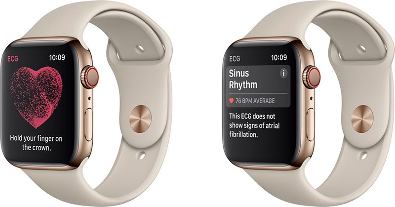
Apple Watch Series 4

"The fear is we could end up with a lot of over-treatment, over-call and people waiting in emergency departments for no good reason."
"[Heart rate variations throughout the day are normal] and now we're going to pick up much more of that signal."
"[A single-lead ECG connection can't diagnose a heart attack] so that's important to get across."
Dr. Samuel Vaillancourt, St.Michael's Hospital, Toronto

"The danger with any new medical technology is that in the enthusiasm to be an early adapter, for what is an admittedly cool gadget, we risk engaging in a population-wide screening strategy that is decidedly flawed."
"[Guidelines don't recommend screening people for AF -- atrial fibrillation], so in a sense no one needs to wear this on the wrist."
"Since the people who are more likely to buy an Apple Watch are younger and probably therefore healthier [wearing an ECG on their wrist] 24/7 is likely going to create a lot of false positives."
"The main risk of AF is that it increases the risk of stroke."
Dr. Christopher Labos, Montreal cardiologist, associate, McGill Office of Science and Society
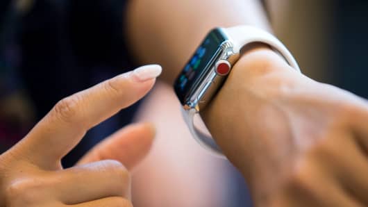
A person uses an Apple Watch Series 3 at the Apple Omotesando store on September 22, 2017 in Tokyo, Japan.
Tomohiro Ohsumi | Getty Images
A person uses an Apple Watch Series 3 at the Apple Omotesando store on September 22, 2017 in Tokyo, Japan

"This is going to create such a massive headache [for doctors]."
"Every worried [healthy] Tom, Dick and Harry are (sic) going to be freaking out about every blip thing that shows up on their Apple Watch."
Dr. Ethan Weiss, associate professor, University of California, San Francisco

"Often patients come to us saying that they feel stuff, but we don't have the rhythm recorded so we can't really tell what's going on."
"I think it behooves us as a medical profession to educate the public that, if you have Afib, it's not the end of the world. [Different if other risk factors for stroke are present, or if long periods of irregular rhythm arise] like days and days, and the heart is really fast."
"[Should the app pick up an abnormality, that should be shared with a doctor for further testing] to figure out if it's atrial fibrillation, or if it's nothing."
Dr. Andrew Ha, cardiac electrophysiologist, Peter Munk Cardiac Centre, University Health Network, Toronto
The people most likely to be or become candidates for atrial fibrillation are the elderly. How comfortable are those in that age category with the use of technologically advanced gadgets that send their younger contemporaries into paroxysms of desire to own the latest, hottest, coolest gadgets on the market? Those already suffering from compromised health conditions and whom readouts of their heart rhythm at any given time presented to their cardiologists for whom the Apple Watch with its over-the-counter ECG could be useful wouldn't be using it; too complex, too confusing.

AF is a leading cause of stroke, affecting approximately 34 million people worldwide. Apple's ECG app is not endorsed by the American Heart Association, but it does admit the device could possibly lead to swifter diagnoses of AF. The recordings, stored in a health app, can generate a PDF to be delivered to the wearer's doctor for consultation, in the best of all possible scenarios. However, given the demographic that will be wearing the watch, it isn't too likely that this scenario will emerge from a gadget that impresses with its capability but in reality is just another plaything.

According to the Apple Watch specifications, the ECG app detects signs of an erratic heart rhythm. But under many circumstances, some normal, some adverse, heart rhythm fluctuates during the space of a day. Separating the wheat from the chaff as it were, is a diagnostician's professional aptitude and concern, not the wearer of the watch who has no medical experience and would be prone to imagining worst-case scenarios unfolding.

So even though the app, according to Apple's description of its function, generates a "heart rhythm classification" with the potential of determining whether heart is beating normally, or whether signs of atrial fibrillation have been detected, what is the wearer going to do with that information aside from panicking? AF causes the heart to quiver and possibly beat chaotically rather than contract in a measured way with each heartbeat. The risk of AF is blood pooling in the heart's upper chamber, where clots can form and move through arteries feeding the brain, to cause a stroke.

A normal ECG has a dozen 'leads' placed on the chest for detection of symptoms, while the Apple ECG app has a single lead. What emerges from that single lead can be very misleading and certainly incomplete. Research studies on systematic screening of large populations for the presence of AF has not proven to be useful, according to Montreal cardiologist Labos, in an article published by McGill University's Office for Science and Society.

False positives are the major concern; false positives where a test indicates abnormality when in fact the results of the test fail to reveal anything abnormal at all, have been common occurrences among the healthier contingent of people tested. Should an individual be tested and found to have AF, the presence of additional risk factors like high blood pressure, diabetes or old age would indicate the use of blood thinners. But not everyone diagnosed with AF requires blood thinners.

And according to Dr. Ha, though the Apple app could provide health professionals with a measure of objective proof of an abnormal heart rhythm, that single lead test should never be considered a substitute for a standard, infinitely more accurate ECG interpreted by an expert. Bearing in mind at the same time that some people with AF have no symptoms whatever, while others are unable to function, continually exhausted.

Clearly, diagnosing such conditions can be complicated requiring medical devices more complex than the Apple app, and the experience leading to related treatment of a specialist. Piggy-backing a medical device onto a technological gadget speaks to the technology-fascinated aficionado's appetite to acquire the latest offering in technological advances and in this instance of health-monitoring and diagnosis, Apple has entered a realm where confusion and complication rule.

It should carry the caution of caveat emptor.

Labels: , , ,

Monday, December 18, 2017

Operating Room Surgical Fires

"What can you say to a patient having a skin lesion excised under monitored anesthesia care [MAC] who suffers severe burns to the neck and face from a surgical-site fire caused by unnecessary supplemental nasal cannula oxygen leaking under drapes and towels into the surgical field where electrocautery was used?"
'Oops!' is clearly insufficient."
Dr. John Eichhorn, University of Kentucky College of Medicine and Medical Center 

"If you want to hold the hospital or medical system accountable, you better have deep pockets."
"Because these injuries are seldom fatal or, in the long term, life altering, they don't get a lot of attention."
"It's human interaction and demands on time over thoroughness and efficiency. These are the things that need to change -- we have a system that is just too busy. There is no system that can operate at 100 percent and not fail."
Darrel Horn, former patient safety investigator, Winnipeg Regional Health Authority

"[For a fire to occur], the three elements of the fire triangle must be present: ignition [heat], fuel and oxygen."
Canadian Medical Protective Association
The last thing anyone would expect when they go into surgery is to catch fire. While rare, surgical fires causing injuries and burns occur in Canada, a new review reports.  Astoria.foto

Undergoing surgery to shrink a tumour mass inside a woman's trachea, the operation went extremely awry when the laser switched on by the operating surgeon caused a sudden flashback and a burst of flames where the laser had lit the inflatable cuff around the breathing tube where anesthetic gases and oxygen inside the windpipe were being delivered, from causing the cuff to deflate, gases to leak and the oxygen to be ignited. This resulted in a rare (for Canada) lawsuit when the patient sued for malpractise.

This was an untowardly dangerous event that should never have happened, but it did. The professional body that defends doctors accused of malpractise, in reviewing 54 cases of surgical fires and burns concluded that many patients came out of this type of unexpected complication during surgery with "scarring, disfigurement and psychological trauma". Some burns were occasioned by surgical equipment or chemicals during surgery.

The woman who suffered an "intratracheal fire" might not be comforted to learn through the inspection by experts that the injuries she sustained partially resulted in all likelihood by the anesthesiologist using 100 percent oxygen rather than the recommended lowest possible concentration, between 30 and 40 percent "to prevent OR fires in this scenario".

Alcohol-based antiseptics used to clean and prepare skin before an incision represent the most flammable of solutions which should be given sufficient drying time before any procedure is commenced. In some scenario the solutions, allowed to pool under the patient rather than being wiped up, present another opportunity for fire to erupt. Until completely dry, prep agents are highly flammable. Alcohol vapours can form, readily ignited by heat or a spark from a cauterizing tool. Alternately, lasers were known to have ignited dry gauzes or sponges placed within the incision site.

Malfunction of lasers, the power level too high or the use of an incorrect type of device represent other issues leading to fire, but half of the fires studied occurred when oxygen concentration was not diluted to the lowest possible level during laser surgery on a patient's head, neck or upper chest. As well -- it was pointed out by experts reviewing the cases -- communication breakdowns during surgery were cited along with delays in diagnosing burns.

The 54 cases all occasioned legal actions and the receipt of complaints to licensing colleges. According to patient safety experts, at least ten times as many additional cases would have occurred nationally. Problems such as these are of magnitudes higher elsewhere around the world and particularly in the United States malpractice lawsuits are infinitely more plentiful. In the U.S., up to 650 operating room fires annually are reported, with the true number much higher, reflecting the lack of mandatory reporting in half of the states.

A patient in Seattle had been awarded $30-million in damages two years ago when an endotracheal tube caught fire inside her throat during surgery to remove polyps on her vocal cords. Post-surgery and fire, she no longer is able to speak or breathe on her own. The U.S. Food and Drug Administration launched a surgical fire prevention initiative in 2011, citing cases including flash fires of an eyelid, a bowel explosion, throat fires and drape and gown fires.

In their document, 'The Patient is on Fire! A Surgical Fires Primer', it was noted that "Most fires in the OR will be either on or in the patient. Fires inside the patient are typically small but can be deadly." Among the 54 cases reviewed in Canada, there were no deaths resulting from the events, but five percent did involve "major, permanent" injuries.

Labels: , ,

Wednesday, August 10, 2016

Chatting With Ludwig

"How people construct sentences, what words they choose to use, and even very microscopic fluctuations in their voice are very indicative of changes to their cognition."
"It [robot attendant] waits for you to talk, and when it figures out you've stopped talking, it sends the audio to speech-recognition [technology]."
"There's a very clear link between someone's expressive ability with language and the progress of Alzheimer's disease, generally. People who work with individuals with Alzheimer's disease ... could use a little bit of extra help, which is what we're trying to build."
Dr. Frank Rudzicz, assistant professor, University of Toronto/scientist, Toronto Rehabilitation Institute
Ernest Doroszuk/Postmedia Network
Ernest Doroszuk/Postmedia Network   Dr. Frank Rudzicz with Ludwig.
"Based on [Ludwig's] analysis, we can tailor-make and customize the care the individual receives."
Isaac Weinroth, executive director, One Kenton Place, Toronto long-term care residence
According to the Alzheimer Society of Canada, 564,000 Canadians live with some form of dementia. This is a figure that will likely rise to 837,000 people afflicted with Alzheimer's or other dementia within the next fifteen years. The society states that after age 65 the chance of an individual suffering from dementia doubles every five years. Memory is the first function to begin to lapse, and after that, verbal language skills.

Dr. Rudzica felt it was time to use his skills in medical engineering and computing to produce a specialty robot whose intervention and usefulness would be invaluable to Alzheimer's care-givers. Five years ago he built a robot almost life-sized with a white case, in appearance resembling an early version of the beloved iPod. This robot was geared to following dementia and Alzheimer's patients in their homes, instructing them in basic tasks.

That robot's programming lacked the capacity to listen to the patients and interpret what they had to say. So he launched into the production of a follow-up robot, reducing the original five-foot size to only two feet, and named it Ludwig, in memory of Ludwig Wittgenstein the Austrian-British philosopher whose special interest was language and the meaning of words.

Ludwig is a new innovation to help in the care of the cognitively impaired. He resembles the physical characteristics of a boy with a high-pitched 'voice', wearing brown, unruly hair. And he is programmed to speak and interact with dementia patients, to ask them leading questions, and then 'consider' their responses, whereupon reports are provided to caregivers.

Ludwig is set to be involved in a practical trial test of its capabilities at One Kenton Place in Toronto.
As the robot stands before a patient, a picture appears on a screen, asking the patient to describe what it is they see before them. Ludwig notes his interpretation of the patient's response; whether they are conversationally engaged, seem happy or anxious, and how they're behaving in comparison to previous similar exchanges.

There are three components whose use is the recording of audio and video resulting from conversations. Ludwig has microphones in his ears, a camera in his eyes, and a sensor in his feet. The patient's gaze is tracked as well as body movement, vocal intonation and word choice. This interchange gives Ludwig the opportunity to assess the cognitive health of a patient.

The potential inherent in this little robot's functioning was a year's investment in planning and construction, all achieved at a building cost of $3,000 to produce Ludwig.

Laura Pedersen/National Post
Laura Pedersen/National Post   Elizabeth Graner, a resident of long-term care home One Kenton Place, talks to Ludwig the robot.
Ludwig's home for the near future and some time after that will be in a common room at the Toronto care residence, where a television, card tables and other socializing apparatuses function to engage the residence's patients. He can be approached by residents on their own initiative to engage with him in casual conversational exchange.

Dr. Rudzicz plans to refine the technical capacity of the robot. To program it more precisely on how it will choose to respond to a patient's answer, to ensure that its assessment of the condition of the patient accurately reflects reality, before it reports back to staff who care for the patients. And bureaucratic regulatory considerations must also be addressed; whether to classify Ludwig as a medical device.

The robot is clearly not a game, but a device with a defined and critical purpose as an aid to caring for cerebrally-impaired patients. A device that is meant to change the game of looking after the needs of such patients.

Laura Pedersen/National Post
Laura Pedersen/National Post    One Kenton Place resident Florence Sherman chats with Ludwig.

Labels: , , ,

Tuesday, August 09, 2016

The ICU Supine Bike

"More and more critical care units are starting to push for early activity, early mobilization, to combat the detrimental effects of being in bed."
Rachel Goard, physiotherapist, Ottawa Hospital
Physiotherapists Josée Lamontagne (left) and Rachel Goard supervised while Respiratory Therapy student Patrick Lapointe demonstrated the specially-designed bicycle at the Ottawa General Campus ICU. courtesy of The Ottawa Hospital
There was a time -- not all that long ago -- when surgical procedures required lengthy hospital stays for recovering patients. They were to be observed by staff while bed-ridden in a hospital setting, to ensure that recovery proceeded as scheduled - or hoped, before they would be released. Even birth deliveries necessitated a week's stay in hospital, fifty years ago. Now? barely a day. Even open-heart surgery currently has doctors ordering that once out of intensive care patients are encouraged to get out of bed briefly to help blood circulation and a more speedy recovery.

Newer surgical techniques such as minimally-invasive surgery made possible with technologically advanced medical instruments and surgical training in the techniques involved are becoming more common, and result in less blood lost, less damage to human tissue, and speedier recovery time. Using of state-of-the-art technology to reduce the damage to human tissue when performing surgery. Rather than making long and deep incisions, for example, the surgeon makes several ¾ inch incisions, then slender tubes called trocars come into use.

The intensive care unit of The Ottawa Hospital is undergoing tests to determine whether critically ill patients, exposed to the opportunity to activate their muscular function while in bed will recover faster, post-surgery. The medium is a specialized bike that can be wheeled up to the patient's bed, to enable patients' feet to produce a cycling motion. The hope is that this will avoid atrophying muscles, a problem of long standing.

Confinement to a bed for prolonged periods of time results in severe muscle weakness. When not used, muscles can atrophy quite quickly. Research indicates that healthy people can lose four to five percent of muscle strength for every week of inactivity. The first ten days, in fact, appears the time when muscle loss is most accelerated. The situation is even worse with the use of mechanical ventilators.

Patients placed on a breathing machine for a week or more will see about 25 percent tending to suffer from intensive care unit-acquired weakness. This debilitating condition can be overcome, but it can take years. Dr. Michelle Kho of McMaster University, the holder of a Canada Research Chair in critical care rehabilitation and knowledge translation is leading a two-year clinical trial among seven medical centres across Ontario, with 60 patient-participants.

The bicycle's screen shows the patient's speed and progress, and a cycling avatar keeps patients motivated. (courtesy of The Ottawa Hospital)
The bicycle’s screen shows the patient’s speed and progress, and a cycling avatar keeps patients motivated. (Courtesy of The Ottawa Hospital)
This will represent the largest study of its kind, examining benefits of in-bed cycling throughout a critical care stay. Other clinical studies established conventional physical therapy like stretching, sitting, standing, walking and light exercise for ICU patients as having value in rehabilitation. Patients are scheduled to 30 minutes daily of in-bed cycling with the use of the RT-300 supine bike, along with routine physiotherapy.

Patients' outcomes will be tracked to compare physical capabilities on discharge as compared to patients in the ICU who have received routine physiotherapy exclusively. Muscle strength is to be assessed, as well as the patients' ability to stand and to walk. The purpose of the study is to fully understand whether early intervention in ICU with daily cycling will produce a swifter recovery.

To be observed as well is whether critically ill ICU patients, many of whom are in pain and deeply sedated, can gain from the supervised use of the cycling machine. Some ICU patients are challenged with conditions such as delirium, obesity and irregularities in heart rhythm and blood pressure. Researchers in 2012 at Johns Hopkins University School of Medicine reported "growing evidence" in support of the idea that critically ill patients benefit from early physiotherapy and exercise, as a safe practice.

Even sedated patients at The Ottawa Hospital will be subject to the developing new protocol of some form of physical activity hastening recovery.  According to physiotherapist Rachel Goard, the bed bike is equipped with a motor capable of aiding pedal movement for critically ill and sedated patients, even when they're not awake and aware.

300 supine bicycle that improves the quality of patient’s lives post ICU stay

Labels: , ,

Sunday, July 03, 2016

Minimally Invasive Surgery

"It [keyhole surgery] reduces the impact on patients' lives."
"It was a light switch [first exposure to keyhold incisions and laproscopic tools]. I went to my chief of surgery and said, 'We need to do this."
"Dr. Poulin always used to say that laparoscopic surgery is surgery of the eyes: it's what you see. If you can't see it, you can't do it, no matter how good you are, no matter how well trained you are."
Dr. Joseph Mamazza, The Ottawa Hospital
Surgery during a minimally invasive surgical procedure in the new, high tech operating room at the Ottawa Hospital. Wayne Cuddington / .
Surgery has steadily moved into the future with the help of imaging technology in the operating theatre. Where new optical instruments have brought successful new operating procedures to the fore. Attaching a micro-camera on the end of a surgical scope represented an especial gift that biomedical engineers blueprinted and produced; a boon to both surgeons and their patients. Doctors are being trained in ever greater numbers across Canada in the new technique.

Where, for example, in surgery around the stomach area, the abdomen is inflated with carbon dioxide, a fibre-optic light and camera are inserted allowing surgeons to operate through keyhole incisions as opposed to large, invasive slashes through the epidermis, musculature, sinew and bone to approach the surgical site. Operating through the keyholes now, surgeons view what is happening in real time on a screen.

With the advent of minimally invasive surgical techniques, miniaturized tools are used, guided by computer-assisted tracking and real-time X-rays to allow surgeons to navigate their way through the body interior. Throughout the process there is little blood, as surgeons face a large television monitor to check their instruments' progress within the human body. A technician in an adjoining room full of computers orchestrates the onscreen show for the surgeons.
Surgery photos during a minimally invasive surgical procedure in the new, high tech operating room at the Ottawa Hospital. The story by Andrew Duffy is about the medical revolution that minimally invasive surgery represents, and how the technique was installed at the Ottawa Hospital during the past decade. Assignment - 123285 (Wayne Cuddington)
An X-ray during surgery. Wayne Cuddington
Laparoscopies have been performed for more than 40 years, in tying off Fallopian tubes in a tubal ligation procedure for the purpose of permanent birth control. The techniques used are similar; the carbon dioxide inflation, the fibre-optic light, a keyhole incision and miniature tools. Over the years the procedure graduated to far more complex surgeries like preventing ruptures of aortic aneurysms, splenectomies  and bariatric bypass surgery.

Patients undergoing these surgeries remain in hospital for a day or two rather than for a more usual open-surgery, post-operative week. Gall bladders can be removed quickly in a cholecystectomy with the use of keyhole incisions and laparoscopic tools. Instead of a 10- to 16- centimetre incision in the abdomen, doctors use four tiny keyholes to remove a patient's gall bladder. From gynaecology to opthalmology and paediatric surgery, minimally invasive surgery has revolutionized operations.

The result is less pain involved with the MIS procedure, shorter hospital stays and improved patient outcomes. No need to cut through muscle, resulting in faster recovery times, and less scar tissue. At The Ottawa Hospital the first high-tech operating room, with three more to follow, custom built for minimally invasive surgery, opened last year. The renovation which cost $9-million included a movable, laser-guided GE Discovery IGS 730, the only one yet in Canada.


Media placeholder
Hovercraft-like tool improves surgery 1:56 

This is a high-tech device that produces high-resolution X-ray images in real time which can be married to CT scans to produce three-dimensional pictures of organs and blood vessels. With MIS there is no need for patients to undergo general anesthesia which is a requirement for open surgery.

Labels: , , ,

Saturday, May 07, 2016

Trust Polarized by Misgivings

"I would not want the reader to go away with the view that surgery is unsafe. I think surgery is safer now than it's ever been. But it's also more complex. We have better clinical outcomes from surgery than we've ever had. We have fewer complications than we've had, but there's room for improvement." 
"One patient is identified for a particular procedure, something changes, the OR [operating room theatre] -- a multiple trauma comes to the hospital -- and things are all re-arranged, and sometimes things get out of sync. It's sometimes possible to get the wrong patient."
"This is happening to good surgeons. [With brain surgery] when complications do occur [for example], they can be serious."
Dr Gordon Wallace, managing director, safe medical care, Canadian Medical Protective Association

"Surgical procedures are complex and may carry significant risks for patients even in the best hands."
Canadian Medical Protective Association [CMPA] review and analysis
Many hospitals have introduced surgical safety checklists that should be followed before a single incision is made; in some provinces, they’re mandatory. Fotolia

A recently-released review of surgeries carried out in Canada through the years 2004-13, involving 1,583 cases of doctors accused of malpractise that were handled by the largest liability insurer for Canadian hospitals and their employees (Healthcare Insurance Reciprocal of Canada) lists communications breakdowns, "absent, sparse or illegible" documentation, and incidents where system safety checks were not followed, as factors helping to contribute to serious surgical misadventures.

The review states that over a million surgical procedures take place annually in Canada. Back in 2004 a Canadian study reached the conclusion that an estimated 70,000 instances of preventable medical errors take place yearly in hospitals, over half of them taking place during surgery. In the United States by contrast, medical error is now recognized as the third-largest cause of death, right behind cancer and heart disease. An American study newly published, estimates that 250,000 Americans die annually as a result of medical care "gone awry".

Researchers from the Johns Hopkins University School of Medicine in Baltimore reporting in the British Medical Journal stated that "Medical error leading to patient death is under-recognized in many other countries, including the United Kingdom and Canada". As though the horrendous toll seen in the United States can be minimized by drawing in similar incidents taking place elsewhere in the developed world where medical science has accelerated its understanding of health issues and formulated best practise surgical procedures as a pioneering force for prolonging human life.

The Canadian analytical report concluded that one-third of the cases taking place in Canada resulted in severe harm to patients, ranging from devastating injuries such as major organ damage, or paralysis, to the hastening of death. Most of these surgeries involved non-cancer, non-trauma surgery where the average patient age was 49, and 76 percent were in relatively good health pre-surgery. An earlier investigation had revealed that a mere fraction of errors are reported by staff on an internal basis. Pointing to an obvious universal propensity to shield the medical community from controversy.

Experts reviewing the cases found fault with the care provided in half of them. The top five sites for surgical error were found to be the uterus, gallbladder, colon, chest or abdomen muscles and breast. Patient harm ranged from death to lacerations, punctures, infections, hemorrhage and burns. Cases involved sponges, rolls or other instruments left in patients, or "wrong surgery", where the wrong patient, the wrong procedure or the wrong body part was involved for 12 percent and 18 percent respectively of CMPA and Healthcare Insurance Reciprocal of Canada cases.

Hospitals are well aware of these unfortunate incidents of surgeries leading to "mishaps", as are their staff. In many hospitals surgical safety checklists have been developed, meant to represent a safety protocol to be followed before surgery is initiated. These checklists have become mandatory in some provinces. First, the patient's name and procedure is confirmed. If an eye, or an organ is to be operated on on the left, then the left side is marked on the patient. Surgical teams are expected to perform rigorous counts to ensure that sponges, needles and instruments entering the operating room are all accounted for.

Handout
Handout An x-ray shows a surgical instrument that was left inside a patient's body after surgery. Research suggests that about 70,000 patients a year experience preventable, serious injury as a result of treatments in Canada.

Wrong-side surgery happens on occasion where the wrong organ is removed. Instances of "retained foreign bodies", which could be towels, packing, needles and sponges left inside a patient in error can occur when prolonged surgeries take place, or when complications set in, like heavy bleeding requiring emergency reaction, according to Dr. Wallace. Neurosurgeons and orthopedic surgeons were identified as the most likely to be involved in surgical incidents; distractions of one kind or another can occur, and people being human, make errors in judgement and process.

"Patient safety incident" terminology

Medical condition
Most unexpected poor clinical outcomes result from the advancement of a medical condition (i.e. the disease process).

Harmful incident from healthcare delivery
Harm results from the care or services provided to the patient.

Recognized risks inherent to investigations and treatments
Most investigations and treatments have inherent risks. Certain complications, adverse reactions or side-effects may occur and are independent of who is providing the care.

Harmful patient safety incident
A patient safety incident that resulted in harm to the patient.

No-harm incident
A patient safety incident that reached a patient but no discernable harm resulted.

Near miss
A patient safety incident that did not reach the patient and therefore no harm results. These have also been called "close calls" or "good catches."

Reportable circumstance
A situation in which there was significant potential for harm, but no patient safety incident occurred. (Specific patient not involved)

The Canadian Medical Protective Association  

Labels: , , ,

Monday, November 09, 2015

Modern Medical Miracles

"Doctors don't want to say that 'there's nothing we can do' and offer palliative care, but sometimes that's the only option. We didn't want to ... give up on our daughter. So we asked the doctors to try anything. We had to do something."
"What is that [the new treatment offered] going to do when bags and bags of chemo haven't worked?"
"I thought it was bad news [when her husband called], but then he said, 'It worked', and I just cried happy tears." 
"I consider ourselves lucky that we were in the right place at the right time to get a vial of these cells. Hopefully Layla will stay well and lots more children can be helped with this new treatment."
Lisa Foley, London, England

"It's very exciting but it is early days. There clearly needs to be long-term clinical studies with more patients [to validate the approach]. We need to understand who responds and who doesn't, and what the long-term results are."
Matt Kaiser, head researcher, Bloodwise, British charity

"We thought that the little bit of liquid in the syringe was nothing [injecting his daughter with a one-millilitre vial of cancer-fighting cells]."
"Even though she is well at the moment, we still don’t know what the future holds." 
"She will still have monthly bone marrow checks for now and might be on some medicines for the rest of her life."
Ashleigh Richards
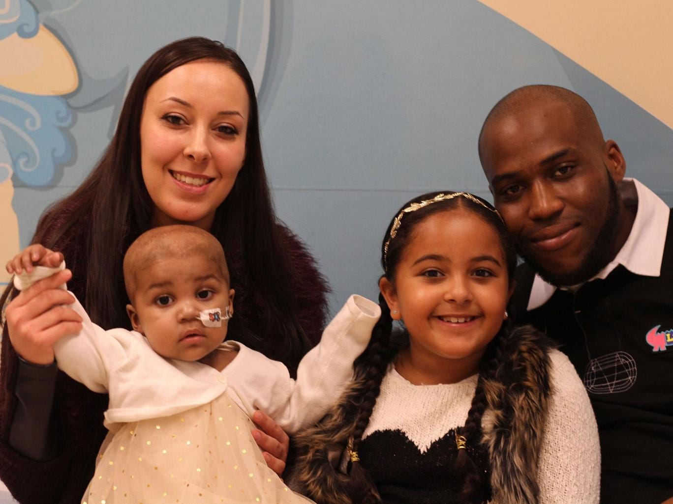
Layla Richards with her parents, Lisa Foley and Ashleigh Richards, and her sister, Reya
Layla Richards with her parents, Lisa Foley and Ashleigh Richards, and her sister, Reya -- PA


"As this was the first time that the treatment had been used, we didn’t know if or when it would work and so we were over the moon when it did. "
“Her leukaemia was so aggressive that such a response is almost a miracle."
Professor Paul Veys, director of bone marrow transplant, Great Ormond Street Hospital, London

"We have only used this treatment on one very strong little girl, and we have to be cautious about claiming that this will be a suitable treatment option for all children."
"But, this is a landmark in the use of new gene-engineering technology and the effects for this child have been staggering. If replicated, it could represent a huge step forward in treating leukaemia and other cancers."
Waseem Qasim, professor of cell and gene therapy, UCL Institute of Child Health
Now a year old, Layla Richard has been given treatment for acute lymphoblastic leukemia since the age of fourteen weeks. She has had dire experiences few children are afflicted with at her young age. Chemotherapy for one, being flown across Britain in vain attempts to determine whether experimental procedures might help, for another. She had bone marrow transplants and innumerable blood tests, all accounting for her one year of life in intensive care units. And the cancer remained.

When her desperate parents, refusing to believe there was nothing more that medical science could offer to save their child's life, pleaded that something, anything more be done, doctors turned to an experimental therapy never before attempted on other than a laboratory mouse. It was not really expected that this new experimental procedure would have an effect on the child's condition and they prepared her parents for that eventuality, even that she might become even more ill.

Layla now is known within the worldwide medical community as the first human being to be treated for cancer with the use of "designer immune cells", as the situation was laid out in an article in Nature, noting the method had previously been tried on HIV patients. And now, Layla has remained cancer free for several months, and the hope is that time will prove her to have been cured by this new serendipitous experimental process.

The treatment itself relates to the editing of genes to prepare the immune cells into which those edited genes where molecular 'scissors' have altered, to be inserted into the cancer patient, they are the body's first line of defence against foreign invasion, which cancer certainly is. The point is to make the immune cells with the edited genes hunt and destroy the cancer. Doctors envision a future where a vial of genetically engineered immune cells would routinely be injected into a patient, resulting in a microscopic war against implacable cancers.

The particular therapy that has proven thus far successful for Layla was developed by researchers at Great Ormond Street Hospital and University College London's Institute of Children's Health, along with the biotech company Cellectis. A bank of pre-engineered, generic T-cells was developed from healthy donors. A genome-editing tool was used to cut the T-cells rendering them impervious to leukemia drugs which would otherwise kill them. New programming directing them to hunt down and fight cancer was pasted into the altered T-cells.

(T-cells are the immune cells that fight off cellular abnormalities and infections, as well as any foreign bodies that the immune cells don't recognize as belonging to the body they protect.) Editing represents a method of prevention against donor cells attacking what else they come across (such as the patient's own cells). The "designer cells" can be contained in vials and shipped out to doctors prepared to inject them into patients whose bodies have been invaded by severe cancers.

It was the first of these "off the shelf" donor T-cells, called UCART19, still in the testing phase, which were injected into Layla. And then the family waited anxiously for results. Nothing appeared to be happening, and the child's parents prepared to spend the last of the time they had left with her before losing her forever. When a rash appeared, it represented a sign Layla's body was responding to the foreign cells infused into her.

Labels: , , , , ,

Sunday, August 30, 2015

Medicine's Astonishing New World

"Ultimately, everyone will want everything 3D-printed. It's not a question of what will be 3D-printed, it's a question of what will not be 3D-printed."
"The ability to work with the [3D-printed model, pre-surgery] model gives you an unprecedented level of reassurance and confidence in the procedure."
"For surgeons who have 3D printing, most won't go into the operating room without it for a complicated procedure."
"It allows for incredible customization. This is the ultimate form of personalized medicine."
"There's great value in going to a place where you can direct a group and develop the technology the way you think it should be done."
"Ultimately, you have to show it's financially viable. But really, I think the savings are going to be unbelievable. It will happen."
Dr. Frank Rybicki, chief of medical imaging, The Ottawa Hospital
Dr. Frank Rybicki holds a 3D model of a child's heart. Models help surgeons plan and perform intricate operations. (Darren Brown/Ottawa Citizen)
Dr. Frank Rybicki holds a 3D model of a child’s heart. Models help surgeons plan and perform 
intricate operations. Darren Brown / Ottawa Citizen
The 3D printer that is on the brink of revolutionizing manufacturing, let alone medical technology, was invented in 1983 by an engineer from an American company that used photopolymers to create a plastic veneer on furniture in their manufacturing technology. During the course of his work, Chuck Hull wondered whether the same protocol used in coating furniture with acrylic-based liquids could be used to produce three-dimensional objects comprised of multiple thin layers of acrylic hardened with UV light from a laser beam.

His first, modest design has long since been improved upon; 3D printers are capable of using all manner of materials to produce the desired object; materials like metals, ceramics, sugar, rubber, plastic, chemicals, wax -- and, amazingly, living cells. The leap from concept and design to the finalized product is amazingly swift. These printers are now indispensable to researchers, to manufacturers, and even to ordinary people wanting to experiment themselves, at home.

As the printers' speed of production has been stepped up, along with their versatility, their cost has diminished, so that even home inventors can acquire these incredible devices. A home desktop version is available from Home Depot for $1,699, and the DaVinci Junior 3D printer is available from Amazon.com for $339.

Medical researchers are now planning to engineer implantable livers, kidneys and other body parts, with 3D printers. New limb joints made from a patient's own tissue and implantable skin for burn victims are being produced by Canadian scientists with the use of 3D bioprinters. Those 3D printers don't, however, come cheap, clocking in at $150,000.

This technology is transforming applications of medical surgeries in ways that could never before be imagined. Doctors in Britain used 3D-printed models, surgical templates and titanium implants to repair facial injuries sustained by a 29-year-old man in a motorcycle accident. A 3D-printed titanium plate solved a hole created by cancer surgery, which caused the patient's eye to sink into the hole when the diseased part of the orbital bone was removed.

Last year in Beijing, surgeons implanted 3D-printed vertebrae in a 12-year-old boy with a malignant tumour in his spinal cord. Doctors in Ann Arbor, Michigan saved a critically ill child by designing and implanting a 3D-printed splint to hold his collapsing windpipe open. Customized body parts, surgical tools, pharmaceutical drugs, and living tissues are now being designed on computers and produced with 3D printers building, layer by layer, three-dimensional objects.

In recent years, 3D printers have succeeded in producing bones, ears, exoskeletons, windpipes, jawbones, cell cultures, stem cells, blood vessels, vascular networks, and organ tissue, according to an article recently published in the medical journal Pharmacy and Therapeutics. Custom-made skull plates, knee implants, hip joints, hearing aids and dentures are now more commonly produced with the aid of 3D printers.

Labels: , ,

Saturday, March 14, 2015

A Unique Mind, An Extraordinary Life

"My father received both the Order of Ontario and the Officer level of the Order of Canada; he received the Living Legend Award from the World Society of Cardio-Thoracic Surgeons. He received Member Emeritus status from the Canadian Medical and Biological Engineering Society -- which gave him particular pleasure because it represented recognition by a college of his peers. And my Dad -- who was one of the founders of the Engineering Institute of Canada -- was named a "Fellow" at last year's awards ceremony."
"My Dad's was a life of dedication, purpose and service ... a life of love of family ... of the essential values and ethics of Judaism, like his parents before him ... of guidance to, and assistance for the betterment of the lives of others. My father embodied Tikkun Olan -- "healing the world" -- and my life will be in permanent deficit by virtue of his absence every day."
Floralove Katz, Ottawa
Leon Katz was made an Officer of the Order of Canada in 2010 by Gov. Gen. Michaelle Jean. Katz died Jan. 9 at age 90.    Chris Mikula / The Ottawa Citizen
What more fitting tribute to a parent, in honour of his dedication to public duty could a proud daughter give than that which this woman wrote in memory of her father's prodigious contribution to making the world a better place? He certainly made the world a better place closer to home for his four children, as he and his wife, their mother, taught them frugality, hard work and respect, the value of education and love of music, conveyed to them both through example and by instruction.

In the public sphere, her father, Leon Katz, born in Montreal in 1924 to impoverished, hard-working people during the Depression years, took his inspiration from his own parents. At the age of 17, Leon Katz volunteered to serve in the Canadian Army. He was a Canadian officer during the Second World War stationed with the British Army, in the Rhine, with 400 Welsh and Scotsmen under his command as he implemented the Military Government Laws.

Later he attended university to graduate as an electrical engineer, and then took another degree in physiology, covering biology, physics and chemistry to lead him to joining electrical engineering with human physiology, heralding the advent, development and advance of biomedical technology and engineering. The end of the Second World War meant that new technologies were released from wartime service; transistors to radioactive isotopes for medical diagnoses.

He excelled in creating original medical devices, teaching their use in clinical practise. And himself taking part in using them in clinical practise. Pairing the industrial and health-care worlds, he created an original contribution of medical devices aiding a large array of medical specialties by conceiving, designing and hand-crafting devices on which he trained scores of others, leading eventually to Canada's first heart-lung pump used in open heart surgery.

In the early 1950s, Leon Katz was one of two biomedical engineers at the Montreal Neurological Institute who provided famed neurosurgeon Dr. Wilder Penfield with instruments, tools and services used in the treatment of brain-related diseases, aiding him in his dramatic discoveries of the human brain. When two doctors at the Montreal Jewish General Hospital in 1953 ventured to explore the potential of I131, the radioactive isotope of iodine in medical applications available from Atomic Energy of Canada's Chalk River reactor, they hired Leon Katz as a physicist.
Radiation physics represented a departure from electrical engineering and neurophysiology, yet Leon Katz organized the first clinical radio-isotope laboratory in the country. He served as founder and director of the Department of Bio-Medical Engineering, Chef, Service de Biophysique; and First Cardio-Pulmonary Bypass Perfusionist at the Institut de Cardiologie, Montreal.

In the 1970s and '80s, Mr. Katz  moved to Ottawa to become the first chief of Diagnostic Devices Division and Evaluation Standards Division, Bureau of Medical Devices in the Health Protection Branch at Health and Welfare Canada. His team there discovered and made correction to the backflow hazard from contaminated evacuated blood collection tubes for the gathering of blood samples.

An article resulting from that finding, published in the distinguished British medical journal The Lancet drew international notice and led to tainted tubes being recalled across Britain and the United States, both countries then following Canada's lead in amending legislation requiring sterilization techniques be implemented to ensure public health standards were upheld.

This amazingly inventive, capable and brilliant mind was finally put to rest when he died in January of 2015 in Ottawa, of mesothelioma and lung cancer, diagnosed only a short time previous to his death at age 90. A man whose life was a tribute to human excellence and altruism.

Labels: , , ,

Tuesday, January 06, 2015

The Good Son

"It's very difficult for me to discuss the facts of the case with him. When I started to read the police report, he just broke down. He put his head into his hands and was sobbing."
"You're 62 years of age. You've never been involved in the criminal justice system. Your mom dies and you're in custody because of this?"
" I can't imagine a more horrible scenario for this fellow."
Mike Cook, lawyer for the defence

"To me it seems like it must have been a misunderstanding."
"Did she really want to lie there in pain for five days, or was it a fear that if you called someone, they would force her to be resuscitated or have care?
"It's absolutely possible to get comfort care (morphine, a catheter, etc.), but not have any treatments that you don't want."
"Care is a broad term, so the education I'd like to give people is to know that you always have the right to refuse a treatment."
"So if she didn't want to go to hospital, [it's] absolutely her right. She didn't want surgery, absolutely her right. But she still could have been given comfort care, and I think had a much more peaceful way to die."
"She clearly wanted comfort care only and wanted to end her life, but she didn't have to do that on a floor, and I suspect in some pain probably from a broken hip."
Wanda Morris, CEO, Dying With Dignity

Ron Siwicki (left) and late mother Betty. Photos from Ron Siwicki's Facebook page and YouTube account.Ron Siwicki (left) and late mother Betty. Photos from Ron Siwicki's Facebook page and YouTube account.


What patience, what devotion from a son to a mother. Repaying, in essence, the mother's investment of love and nurturance for a child that is her son. Of course, with mother at age 89 and son aged 62, though the mother-son relationship remains intact, the son is no longer the child once so dependent on his mother's attention. The positions reverse themselves. With the son devotedly living with his mother, it is the mother who then requires at her advanced age and health disabilities, including dementia, the attention and compassionate care given her.

Such a primary relationship. It is admirable, quite, that a 62-year-old would fashion his life around the care and comfort of a mother in her extreme elderly condition of failing health and advance toward the inevitable. Friends of Ron Siwicki of a Winnipeg, Manitoba address attest to his concerns for his mother's well-being, a dutiful son. He "always carried out his mother's wishes", said one long-time friend.

"Perhaps this was not a wise thing to do here", added Henry Kreindler, also of Winnipeg, in considering the situation that Ron Siwicki has got himself into, and the illegal criminal action he is held to have taken in his obedience to his mother's wishes. Betty Siwicki, mother of Ron, is no longer. She died a painfully excruciating death, refusing care, insisting that she be allowed to finally die, that no measures be taken to prolong a life she clearly meant to leave.

In refusing any kind of medical care that would prolong the life that Betty Siwicki was denying, she was in her legal right under Canadian law. But Canadian law does not state that people intending to die, refusing extraordinary treatment to prolong encroaching death, must die unattended by medical professionals, in pain and suffering before death finally makes its dilatory visit to release her from life's cruel grip.

In her dying days and hours Betty Siwicki could and should have been offered palliative care. The medical community is prepared, as it should be, to proffer that compassionate end as a basic human right. Betty Siwicki had no intention of being subjected to end-of-life life-support. By law doctors cannot insist on providing invasive care if the patient has no wish to receive it. To do so would be to open themselves to potential assault charges.

Betty Siwicki collapsed in her bedroom and lay there, injured and in pain on the floor for five days, with her son obedient to her wishes, leaving her there, but thoughtfully providing the blanket she requested. He also gave her protein drinks on occasion, at her request. Obviously among his other traits, he is a man of patience, and he waited as she bid him, to allow her to die. Dignity, obviously had no part in this very intimate drama.

On her death, Ron Siwicki, the bereft son, was arrested. And charged with criminal negligence causing death and failing to provide the necessities of life. One of the charges is more commonly reflected by situations of extreme child neglect, the other by situations of criminal neglect; like drinking and driving. After all, Ron Siwicki chose to be obedient to a mother not quite capable at that point of making logical decisions yet he abided by her rule, though he need not have, and spared her the agony she suffered before dying.

Perhaps this was indeed not a wise thing to do.

Labels: , , , ,

Saturday, August 30, 2014

Understanding Cancer

"At an airplane terminal, you have all of your paperwork in place and there are guards to check it and make sure you're secure. The body is the same way."
"The immune system checks cells as they escape and filters them. This process of escape from the bloodstream is an important checkpoint where most of the cancer cells are destroyed. But if they're able to produce these invadopodia -- the right paperwork -- they're able to escape."
"No man will die of prostate cancer if it stays in his prostate. It becomes dangerous when it spreads. The prostate is not life-threatening if you lose it."
Dr. John Lewis, associate professor, University of Alberta, department of oncology
Cancer cells are cells that grow and divide at an unregulated, quickened pace. Although cancer cells can be quite common in a person they are only malignant when the other cells (particularly natural killer cells) fail to recognize and/or destroy them.[1] In the past a common belief was that cancer cells failed to be recognized and destroyed because of a weakness in the immune system. However more recent research has shown that the failure to recognize cancer cells is caused by the lack of particular co-stimulated molecules that aid in the way antigens react with lymphocytes.[2
Dr. Lewis must know of what he speaks, with such ease and familiarity. He is, after all, the holder of the Frank and Carla Sojonky Chair in Prostate Cancer Research at the university. And knowledge about how cancer spreads is of huge importance. So it can be detected, prevented if at all possible, and if not, how to manage it before it becomes too late.

For example, research conducted by the university research team of which Dr. Lewis is an integral part, found that doctors could use drugs or genetic means to halt the development of invadopodia.

And what in the name of heaven is invadopodia?!
Figure thumbnail fx1

It is described as cancer cells forming "tentacles" enabling the cancer to spread from one body part to another. Those tentacles are invadopodia...makes sense doesn't it? It took three years of intense observation to discover how cancer cells the size of microns developed invadopidia, allowing them to move within the bloodstream to invade another organ. This is a phenomenon never before observed in a live model.

And, as Dr. Lewis said, the deadliest thing about cancer is its spread to other organs in the body. The spread of cancer, called metastasis is the cause of death for 90% of cancer patients. The diagnosis of metastatis is recognized by most people as a death sentence, often sooner than later.  There is now evidence that performing a biopsy or surgery on a cancerous tumour can cause cancer to spread.

In which instance an invadopodia inhibitor drug would be particularly important to use in such cases.

The drug that was used by the University of Alberta research team is now in clinical cancer trials; "encouraging", comments Dr. Lewis. He and his team of researchers made use of a $500,000 microscope and the protein of a deepsea jellyfish for their work. The protein glows fluorescent green, showing clearly in images of the cancer cell on a blood-red backdrop.

U of A scientist puts a finger on cancer cell ‘tentacles’; New study looks at how cancer spreads through the body
A University of Alberta research group injected fluorescent cancer cells into the bloodstream of test modules, then used high-resolution, time-lapse imaging to see what happens to them. Photo: Supplied


Purpose-built for the study, it is one of only two such microscopes in the world. The Alberta Cancer Foundation helped to fund the microscope and the study, and helped to bring Dr. Lewis's expertise from Ontario's Western University to Alberta, and the University of Alberta.

The team’s work was published in the most recent issue of the journal Cell Reports. Some of the study work was done by scientists at the Lawson Health Research Institute in Ontario.


Labels: , , , , ,

Thursday, July 03, 2014

Lean Right In To LEO

Don't we love electronic gadgets. And aren't we infused with enthusiasm over fitness, becoming fit, staying that way. And then, before all of that, there is the fixation on dieting and losing weight. Put them all together and you have the perfect storm of intention prodding energy to be directed toward:
  1. Losing weight;
  2. Using a gadget;
  3. Working out.
"I lost half my body weight. I looked like a stick. I started lifting weights, and I realized that there is no product out there that allows you to measure muscle activity and growth. It just didn't exist. Certainly not like there is for measuring heart rate and calories."
" In 2012 I had some great students come along. We really got serious about it and we built a fully functional electromyographical mobile system. This is something you can wear and walk around and use."
"For example, when you're cycling, you are supposed to pull with your hamstrings."
"We even came to the idea that we could use it [the LEO] for injury prevention. Most injuries in cycling and running are self-induced."
Leonard MacEachern, professor, department of electronics, Carleton University, Ottawa

Just as well Dr. MacEachern was in the university's department of electronics. He was using heart-rate monitors in his weight loss program, and thought there must be a similar electronic device for the purpose of monitoring muscle contractions, lactic acid buildup and other indicators to assist in furthering knowledge about just how successful his exercise program was, and to inform him whether or not he was actually doing things properly. A device that might warn him whether he might be causing injury to himself.

But he discovered soon enough that there was nothing out there to fit the bill, that technology hadn't yet caught up with the need for people exercising to monitor the key components of their body functions and reactions to exercise regimens and possibly undue stress they might be unaware of. Having that information available to them on such a personal level could enable them to correct deficiencies and re-direct their movements while at the same time alerting to the potential of self-harm.

GestureLogic: Wearable computing monitors athletes' muscles
Engineering professor Leonard MacEachern of Carleton University, along with his team of students, has developed technology that by using a band similar to a tensor bandage only with high tech sensors and worn on the thigh while cycling or running, an athlete can reduce injury by being able to track things like lactic acid build up etc. They can then adjust their work out. Photo taken at 13:51 on June 25, 2014. (Photo by Wayne Cuddington / Ottawa Citizen)

With the help of some of his students exercising their creative abilities and imagination the result was a product that he and his team named LEO. It is a device anchored within a band constructed of conductive fibres wrapping around an individual's upper thigh which then relays data to a personal device like an iPhone or Android device. That information captured by LEO renders details such as which muscles in the thigh are firing, how emphatically, the distance being covered, how much lactic acid builds in the muscle; heart rate, and energy burned.

This kind of contraction monitoring, lactic-acid buildup and energy expended enables LEO to warn an athlete when an activity, or the manner in which it is performed may result in an injury. GestureLogic, the company that Mr. MacEachern built after six years of technological manipulation perfecting a wearable computing device to map muscle contractions to produce LEO, plans to target the device initially to the running and cycling crowd.
LEO
The Ottawa-designed and produced device that GestureLogic with its 11 employees is now prepared to market plans to bring it to a crowdfunding platform on Indiegogo. One of any number of products identified as "wearable computers", and for which the takeup among gadget and health-conscious consumers has enormous potential, LEO has high hopes for an initial sales impact. Hoping to raise at least $50,000 to set GestureLogic on the right sales track through the Indiegogo campaign the company plans to offer LEO for a discounted $179 during the campaign.

After which LEO's recommended retail sales price would rise to $299. The experience gained by the company through its social fundraising campaign will help Mr. MacEachern in setting future pricing and identifying marketing device initiatives. But according to technology researcher BCC Research, the market for wearable computing devices is set to soar.

Labels: , , ,

 
()() Follow @rheytah Tweet